
Know Your joints
Know Your joints


- The joint is primarily formed by the two large bones of the lower limb, the Femur (thigh bone) and the Tibia (shin bone). The Patella (kneecap) articulates with the femur at the front of the knee.
- The fibula joins with the tibia on the lateral (outside) side of the knee. Together, the femur, tibia, and patella make three compartments (Medial, Lateral, and Patellofemoral).
- Each of the bones has a bearing surface of articular or hyaline cartilage.
- The meniscus in each of the medial and lateral compartments. The menisci are like cushions or spacers and are made of fibrocartilage.
Knee is divided into three major compartments:
- Medial compartment (the inside part of the knee)
- Lateral compartment (the outside part)
- Patellofemoral compartment (the front of the knee between the kneecap and thighbone)

The hip joint is a Diarthroidal Joint whose unique anatomy enables it to be both extremely strong and amazingly flexible, so it can bear weight and allow for a wide range of movements. The primary function of the hip joint is to provide dynamic support to the weight of the body/trunk while facilitating force and load transmission from the axial skeleton to the lower extremities, allowing for ambulatory and mobility functions.
The hip is located where the head of the femur, or thighbone, fits into a rounded socket (Acetabulum)of the pelvis. This ball-and-socket construction allows for three distinct types of flexibility:
- Hip flexion and extension – moving the leg back and forth;
- Hip abduction and adduction – moving the leg out to the side (abduction) and inward toward the other leg (adduction); and
- Rotation – pointing toes inward (internal rotation) or outward (external rotation) and then moving the straightened leg in the direction of the toes
- Hip bones, including the femur and pelvic bones;
- Hip articular cartilage – Labrum rings the outer edge of the acetabulum. The labrum deepens the socket joint, making the joint more stable, but its elasticity allows for flexibility and decreases friction between the bones and allows for a smooth gliding motion
- Hip muscles that both support the joint and enable movement; The groups of muscles supporting the hip are gluteal muscles, located on the back of the hip (buttocks);
- The adductor muscle on the inner thigh;
- The iliopsoas muscle, which extends from the lower back to upper femur;
- Quadriceps, a group of four muscles that comprise the front of the thigh; and
- Hamstrings, a group of muscles that comprise the back of the thigh and extend to just below the knee
- Hip ligaments and tendons, tough, fibrous tissues that bind bones to bones and muscles to bones; and
- Synovial membrane and fluid: A synovial membrane encapsulates the entire hip joint. This membrane produces synovial fluid, a viscous substance that lubricates and circulates nutrients to the joint.
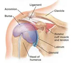
The shoulder joint is among the most flexible joints in your body. It is a ball-and-socket joint and is made up of three bones: the upper arm bone (humerus), shoulder blade (scapula), and collarbone (clavicle).
The ball at the top end of the arm bone fits into the small socket ‘glenoid’ of the shoulder blade to form the shoulder’s glenohumeral joint. This is surrounded by the soft tissue labrum. The articular cartilage which is a smooth, durable surface on the head of the arm bone, along with the thin inner lining synovium of the joint allows the smooth motion of the shoulder.
The shoulder joint also comprises a thin sheet of fibers called capsule that surrounds the shoulder joint, allowing a wide range of motion, yet provides stability. A rotator cuff is a group of muscles and tendons that attach your upper arm to your shoulder blade and is an integral part of the joint.
The rotator cuff covers the shoulder joint and joint capsule. The muscles attached to the rotator cuff enable one to move and rotate their arms in multiple directions such as front, above, to the side, and behind your body.
But with this flexibility, the shoulder also becomes vulnerable to instability and injury. Timely and proper diagnosis helps avoid severe repercussions later and treat the joint before the condition worsens. If the complication or ailment in the shoulder is quite serious, then the Joint Replacement Surgeon may go for open shoulder surgery instead of arthroscopy.

The elbow is the synovial joint between the upper and lower parts of the arm. It is the point of articulation of three bones: the humerus of the arm and the radius and the ulna of the forearm. These bones give rise to two joints:
- The humeroulnar joint is the joint between the trochlea on the medial aspect of the distal end of the humerus and the trochlear notch on the proximal ulna.
- The humeroradial joint is the joint between the capitulum on the lateral aspect of the distal end of the humerus with the head of the radius.
The elbow forms from the expansion of the lower end of the humerus into two thick knobs, or condyles: the humerus’ dome-shaped lateral condyle articulates with a shallow depression on the end of the radius, and the humerus’ spool-shaped trochlea fits into a notch in the ulna. In addition to this, the edge of the radius’s head fits into a shallow groove on the side of the ulna. The bending and extension of the elbow joint are achieved, respectively, by contractions of the biceps and triceps muscles. These movements chiefly involve only the humerus and ulna; rotation of the forearm involves the smaller radius bone as well.
There is a collection of ligaments that connect the bones forming the elbow joint to each other, which aid in the stability of the joint. The humeroulnar and the humeroradial joints each have a ligament connecting the two bones involved in the articulation: the ulnar collateral and the radial collateral ligaments.
Similarly, like almost all other synovial joints, there is a thin layer of smooth articular cartilage that covers the ends of the bones that form the elbow joint. The joint capsule of the elbow surrounds the joint to provide strength and lubrication to the elbow. Slick synovial fluid produced by the synovial membrane of the joint capsule fills the hollow space between the bones and lubricates the joint to reduce friction and wear.
Often one can experience wear and tear in the elbow joint and it is a common site for injury. In such cases, orthopedic treatments may be required to cure and treat elbow joint issues.
Meet Our Doctor

Dr. Bhavya Shah
MBBS, MS ORTHOPAEDICS,
FELLOWSHIP IN JOINT REPLACEMENT SURGERY,
FELLOWSHIP IN PELVI-ACETABULAR TRAUMA AND COMPLEX TRAUMA SURGERIES
Dr. Bhavya Shah is Founder ofNavkar Orthopaedic Clinicone of the leading orthopaedic surgeon in Ahmedabad.